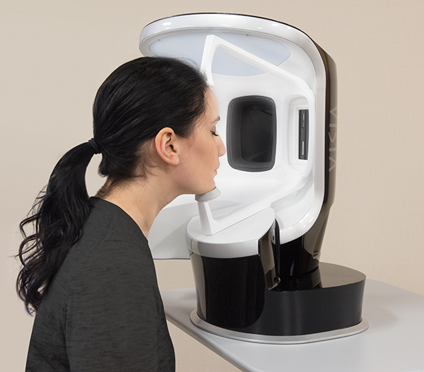
Here at Inject, we strive to stay up to date on the latest technologies with research-based evidence, which is why we are excited to bring on the Visia Skin Analysis!
Here at Inject, we strive to stay up to date on the latest technologies with research-based evidence, which is why we are excited to bring on the Visia Skin Analysis!
Visia uses IntelliFlash®, cross-polarized and UV lighting, used to record and measure surface and subsurface skin conditions. UV photography provides the most complete data set available for sun damage assessment and analysis, including UV fluorescence imaging to reveal porphyrins.
Canfield’s RBX® Technology separates the unique color signatures of Red and Brown skin components for unequaled visualization of conditions that result in color concentration, such as spider veins, hyperpigmentation, inflammation and other conditions.
Your Visia Scan Analysis will capture left, right and frontal facial views (before your scan, you will be asked to wash your face in office. Your scans will be the most accurate with a clean and dry face!).
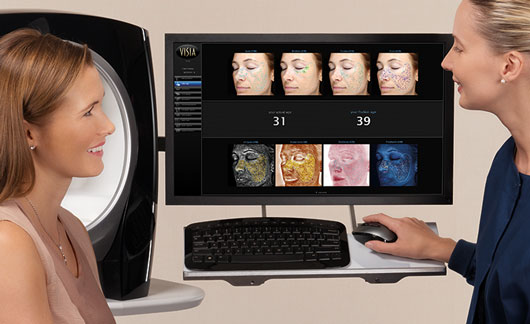
After capturing those views, a quantitative analysis & visual assessment with multiple images will be available immediately to review. Your scans will show:
Spots:
Spots are typically brown or red skin lesions including freckles, acne scars, hyper-
pigmentation and vascular lesions. Spots are distinguishable by their distinct color and contrast
from the background skin tone. Spots vary in size and generally have a circular shape.
Pores:
Pores are the circular surface openings of sweat gland ducts. Due to shadowing, pores
appear darker than the surrounding skin tone and are identified by their darker color and circular shape. The VISIA system distinguishes pores from spots based on size; by definition, the area of a pore is much smaller than a spot.
Wrinkles:
Wrinkles are furrows, folds or creases in the skin, which increase in occurrence as a
result of sun exposure and are associated with decreasing skin elasticity. This skin feature has the greatest variability from image to image as it is highly dependent upon the facial expression of the client. Wrinkles are identified by their characteristic long, narrow shape.
Texture:
Texture is primarily an analysis of skin smoothness. Texture measures skin color and
smoothness by identifying gradations in color from the surrounding skin tone, as well as peaks (shown in yellow) and valleys (shown in blue) on the skin surface that indicate variations in the surface texture.
Porphyrins:
Porphyrins are bacterial excretions that can become lodged in pores and lead to acne. Porphyrins fluoresce in UV light and exhibit circular white spot characteristics.
UV Spots:
UV spots occur when melanin coagulates below the skin surface as a result of sun damage. UV spots are generally invisible under normal lighting conditions. The selective absorption of the UV light by the epidermal melanin enhances its display and detection by VISIA.
Red Areas:
Red Areas represent a potential variety of conditions, such as acne, inflammation, Rosacea or spider veins. Blood vessels and hemoglobin contained in the papillary dermis, a sub-layer of skin, give these structures their red color, which is detected by the RBX Technology in VISIA. Acne spots and inflammation vary in size but are generally round in shape. Rosacea is usually larger and diffuse compared to acne, and spider veins usually are short, thin and can be interconnected in a dense network.
Brown spots:
Brown Spots are lesions on the skin such as hyper-pigmentation, freckles, lentigines, and melasma. Brown Spots occur from an excess of Melanin. Melanin is produced by melanocytes in the bottom layer of the epidermis. Brown Spots produce an uneven appearance to the skin and are detected in VISIA by RBX.
Based upon your scans, personalized recommendations from our built-in library of skin care products and treatments will be discussed with your provider. With ViewMyConsult, you will be able to view your pictures, treatment recommendations, and treatment progress at home using a secure, password-protected web portal. Printed reports are also available!
For more questions or to schedule your Visia scan, please call or text 918-712-1767.
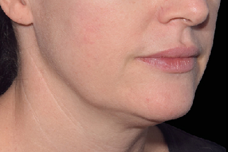
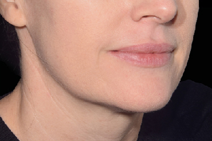
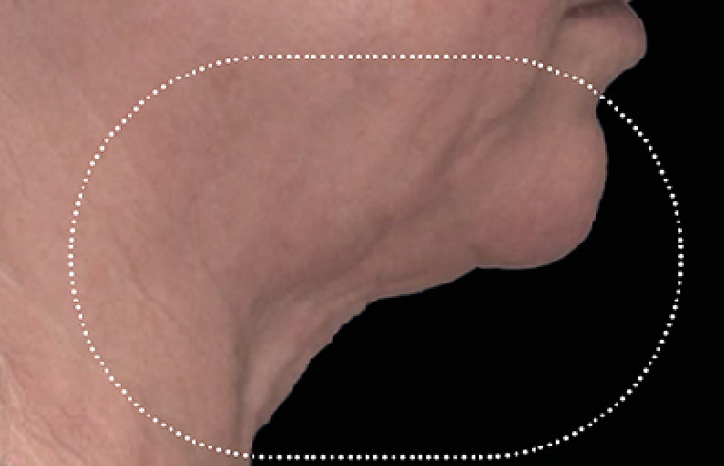
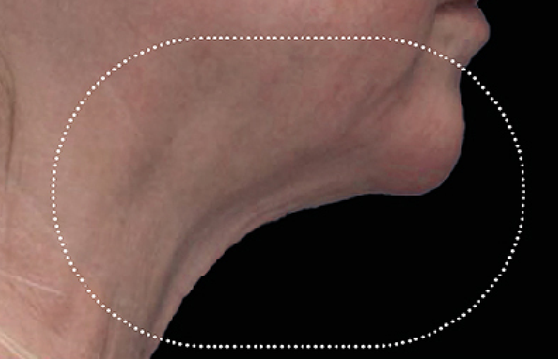
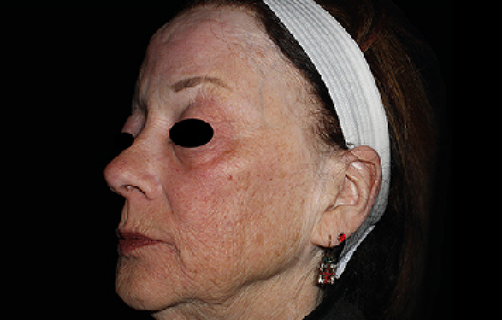
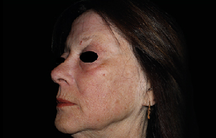
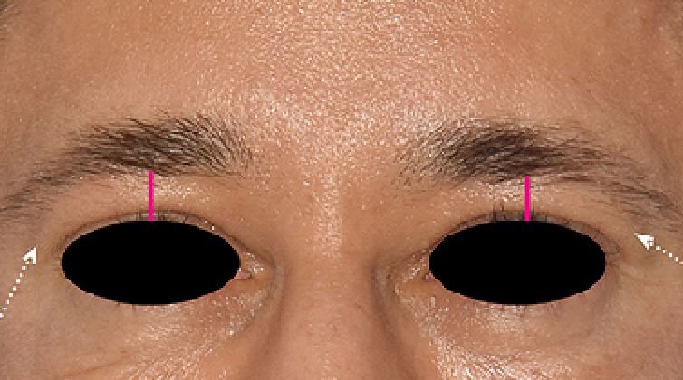
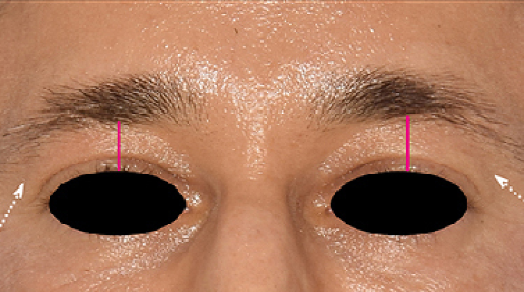
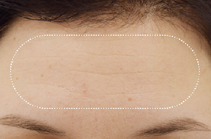
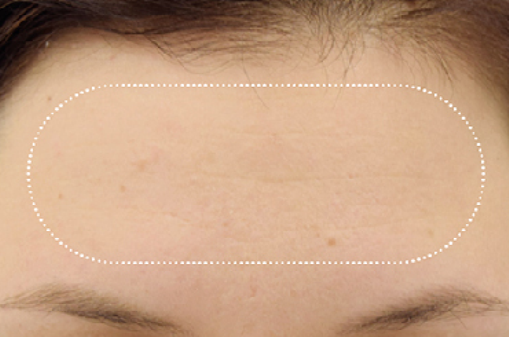
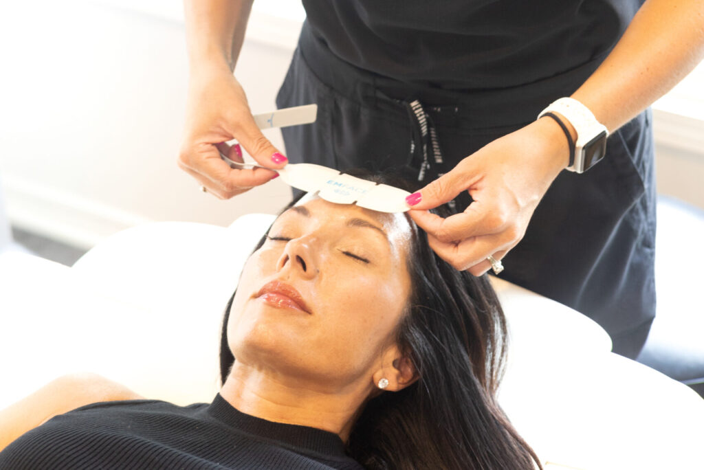
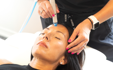
Discover Your Potential: Explore Before and After Photos to Visualize the Incredible Enhancements Achieved with EMFACE—Your Pathway to Radiant Confidence.
EMFACE combines RF (Radiofrequency) technology and HIFES (High-Intensity Focused Electromagnetic) energy to stimulate facial muscles and increase collagen production, resulting in tightened skin and improved facial contouring.
EMFACE treatment is generally well-tolerated by most individuals and is not considered painful. Some patients may experience mild discomfort or a tingling sensation during the procedure.
While results may vary depending on individual factors such as skin condition and treatment goals, many patients begin to notice improvements after just one session. A series of treatments spaced several weeks apart is often recommended for optimal results.
EMFACE treatment is non-invasive and typically associated with minimal side effects. Some patients may experience temporary redness, swelling, or mild discomfort in the treated area, which usually resolves within a few hours to days.
EMFACE is suitable for individuals seeking non-surgical facial rejuvenation and improvement in facial symmetry. It is generally safe for most skin types and tones. However, a consultation with a qualified healthcare provider is recommended to determine candidacy and develop a personalized treatment plan.
COMBINE
WITH
Elevate Your EMFACE Experience with Supporting Treatments. Unleash Your Skin’s Radiance with Our Combined Services.